
MyoTak
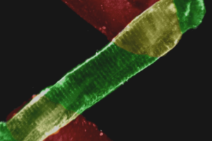
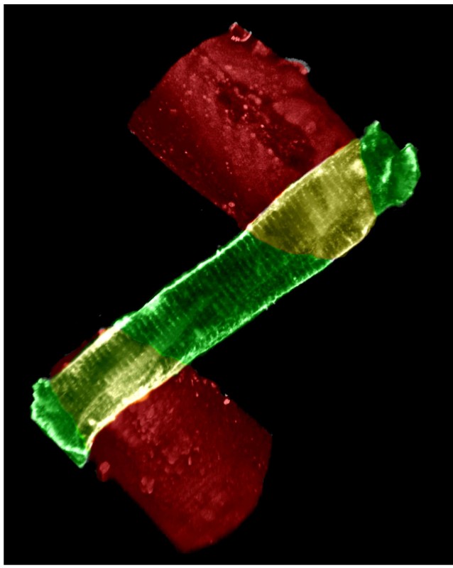
IonOptix MyoTak is a specially-formulated biocompatible adhesive that permits the attachment of cells to surfaces that are smooth or otherwise difficult to adhere to. Developed and patented by researchers at the University of Maryland, IonOptix MyoTak provides a bond strong enough to allow direct force measurements from single primary cardiac myocytes yet is non-cytotoxic and will not damage cell membranes or otherwise affect cellular physiology (B. Prosser, C. Ward and W.J. Lederer, Science 2011). IonOptix MyoTak will readily coat a variety of materials including carbon fibers (P. de Tombe, unpublished data) as well as small glass rods (B. Prosser, C. Ward and W.J. Lederer, Science 2011).
IonOptix MyoTak can be used to attach cells for a number of purposes, including:
IonOptix MyoTak consists of two components: a Pre-Coat and Glue. The IonOptix MyoTak Pre-Coat is used to prepare surfaces for application of the IonOptix MyoTak Glue, while the Glue enables direct attachment to cell surfaces. We are proud to offer IonOptix MyoTak to the scientific community.
Note: This product is intended for research purposes only. It is not certified for clinical applications (including diagnostic purposes). Use of this product in uncertified applications is in violation of FDA regulations.