Go back to RESOURCES
Pressurized Blood Vessel


Interest in the physiology and pathology of the circulatory system has led to a substantial increase in the number of laboratories studying isolated and pressurized blood vessels. Although the relationship between intracellular second messengers and vascular constriction/ dilation has been an area of intense investigation for decades, elucidation of the fundamental molecular and biophysical mechanisms remains at the forefront of vascular physiology research. Impairment to the regulatory machinery governing vasodilation and vasoconstriction correlates strongly with the onset and progression of many pathologies, including cardiomyopathies. Arterial diameter, flow and calcium recordings from arterial segments offer an important physiologic measurement, providing key insights into the processes that influence vascular health.
IonOptix developed its Vessel Recording Systems over many years of collaboration with top vascular researchers. We take pride in a line of precision products that are application driven and built to meet the needs of a demanding research environment. Since its inception in 1990 IonOptix has built and installed hundreds of high performance, turnkey systems in research laboratories worldwide.
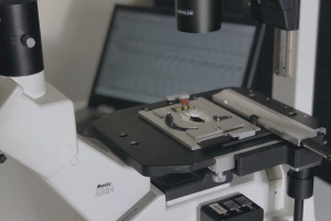
Pressure myographs are used to cannulate and pressurize small arteries in a controlled manner. Placing the myograph on a microscope allows the outer and luminal diameters to be tracked while also measuring fluorescence signals from the myograph. When combined with your pressure myography system, our vessel systems provide everything necessary for simultaneously acquiring vessel calcium, flow and geometry data with our IonWizard 6 core software and VesAcq, FloAcq and PMTAcq acquisition modules. Our complete systems include an inverted fluorescence microscope equipped with a calcium photometry objective and brilliant optics, a digital dimensioning camera and a suite of analog and digital connections for synchronous data collection.
Diameter The IonOptix VesAcq acquisition module tracks vessel geometry, including outer and luminal diameters, wall thickness, cross-sectional area and Media/lumen ratio. Advanced edge detection algorithms applied to the contrast information within digitized images allow dynamic tracking of vessel edges. A new, simplified interface improves the reliability and reproducibility of the tracking and measurements. Data are displayed in real time so that the investigator has immediate feedback.
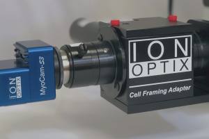
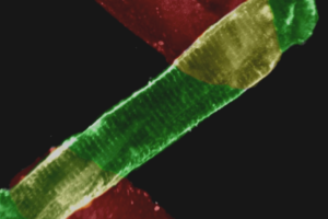
Fluorescence IonOptix specializes in measuring ratiometric fluorescence, which is used to quantify intracellular Ca++, H+(pH) or Na+ concentrations, but the system is also suitable for general quantitative fluorescence and advanced techniques such as FRET. IonOptix provides all the necessary hardware for rapid wavelength switching, coupling fluorescence excitation light into the microscope and detecting and quantifying the emitted fluorescence.
Flow IonOptix’ FloAcq acquisition and recording module provides real-time indicators of arterial flow dynamics, based on raw flow rate and inlet and outlet pressure inputs from your myography system along with dimensioning outputs from our VesAcq software. Output data include mean pressure, flow velocity, wall shear stress – a characterization of the frictional drag exerted on arterial walls during flow, vascular resistance – a definition of the force opposing the movement of solution through a vessel, and Reynolds number – a measure of flow stability and turbulence. Click here for more information on vessel flow characterization.
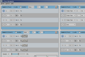
Myographs To be able to deliver fully integrated systems, we have adapted our hardware and software to work together with the pressure myographs from the two leading manufacturers, Danish Myotechnology (DMT) or Living Systems (LSI). Our signal generator (a core IonWizard 6.1 feature) offers the ability to drive external devices such as pressure interfaces.
Below are links to some relevant papers that are published with combined IonOptix / pressurized myograph systems. It also provides the links to the publication abstracts.