Go back to RESOURCES
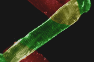
This article, published in Cells, demonstrates the utility of human cardiac organoids (hCOs) to model hypertrophic cardiomyopathy (HCM) pathogenesis. hCOs were generated from hiPSC-CMs from either noncarrier or D389V carrier human subjects. Following maturation into 3D organoids, functional studies were performed. The D389V hCOs showed significantly faster contractility kinetics that could be reversed by treatment with the myosin inhibitor mavacamten. Similarly, D389V hCOs showed faster calcium handling. Additionally, RNAseq analysis showed upregulation of contractility-associated genes as well as genes associated with oxidative stress in D389V versus noncarrier hCOs.
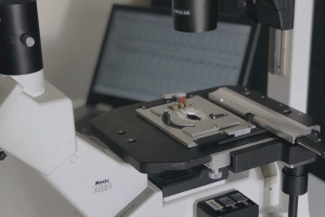
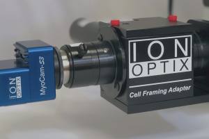
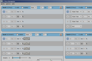
For calcium and contractility data acquisition and analysis, the authors of this paper utilized their IonOptix CytoCypher MultiCell System equipped with CytoMotion for hCO contractility measurements and CytoSolver for analysis (see paper supplementary materials for expanded methods). Please contact us if you’re interested in performing similar functional measurements in cardiac organoids in your lab.
Desai D, Song T, Singh RR, Baby A, McNamara J, Green LC, Nabavizadeh P, Ericksen M, Bazrafshan S, Natesan S, Sadayappan S. MYBPC3 D389V Variant Induces Hypercontractility in Cardiac Organoids. Cells. 2024 Nov 19;13(22):1913. doi: 10.3390/cells13221913.