Go back to RESOURCES
In this recent paper published in Nature Scientific Reports, Researchers at University and University Hospital Würzburg, Germany, develop a novel workflow to characterize and quantify both intracellular and extracellular cardiac sodium levels ([Na+]i and [Na+]e, respectively) and to investigate Na+ homoeostasis in cardiac dysfunction and failure.
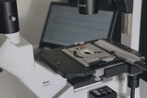
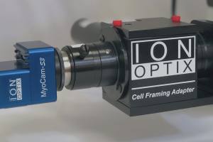
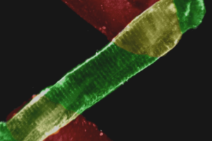
For [Na+]i measurements, the researchers used their IonOptix MultiCell microscope to quantify the ratiometric fluorescent indicator sodium-binding benzofuran isophthalate (SBFI). To derive [Na+]e, they used a multi-compartment model combining [Na+]i with Na+ magnetic resonance imaging to quantify tissue sodium concentration and extracellular volume noninvasively. The investigators then went on to demonstrate the utility of their approach by characterizing Na+ storage in disease-model mice. Interestingly, intracellular and extracelluar Na+ distributions differed among mice that underwent trans-aortic constriction (hypertrophic model) vs. those that underwent left anterior descending artery ligation (myocardial infarction model).
IonOptix got its start in 1990 by offering fast fluorescence data acquisition, and it’s been one of our core capabilities ever since. If you’re interested in performing quantitative fluorescence measurements, contact us and let us know. Whether it’s our MultiCell, standard Calcium & Contractility, Cardiac Slice, or any of our other instruments, we’d be happy to configure a system to meet the specific needs of your investigations.
Christa M, Dithmar F, Weinaus T, Kohlhaas M, Arias-Loza AP, Hofmann M, Elabyad IA, Gutjahr FT, Maack C, Bauer WR. A new approach to characterize cardiac sodium storage by combining fluorescence photometry and magnetic resonance imaging in small animal research. Sci Rep. 2024 Jan 29;14(1):2426. doi: 10.1038/s41598-024-52377-w.