US: +1 617 696-7335 | EU: +31 317 728 104|info@ionoptix.com
  • IonOptix HyperSwitch

HyperSwitch Light Source

2019-11-11T10:34:10-05:00

HyperSwitch Light Source In cases where highest time resolution is essential, the IonOptix HyperSwitch Light Source is recommended. Wavelength switching is accomplished in less than 1msec and can be continuously alternated at rates as high as 250 pairs/sec. The HyperSwitch can also produce hardware video-locked wavelength switching. This figure illustrates the optical path and components of the IonOptix HyperSwitch Dual Excitation Light Source. The light output of a [...]

HyperSwitch Light Source2019-11-11T10:34:10-05:00
  • IonOptix Intact Muscle Chamber

Intact Muscle Chamber

2019-11-11T10:27:34-05:00

Interested in a COMPLETE Intact Muscle Chamber system? Learn more here Lying between single cell and whole heart experimental studies, intact papillary muscle isolated from hearts offers unique advantages. Unlike single isolated myocytes, which represent the smallest fully functional model system, intact papillary muscle can be used to study cardiac function within a multicellular context and an intact 3-dimensional myofilament lattice. And unlike whole heart studies, the contractile characteristics of papillary [...]

Intact Muscle Chamber2019-11-11T10:27:34-05:00
  • LED light source

LED Light Source

2024-02-01T16:37:50-05:00

LED Light Source Recent advances in light emitting diodes have led to a number or previously unavailable wavelengths, including higher energy UV LEDs for Fura-2 illumination. We're proud to offer advanced LEDs offering excitation light for most common fluorophores. These LEDs feature closed-loop feedback control for stable intensity as well as protective overload circuitry to prevent LED damage. Wavelength switching is practically instantaneous and can be continuously alternated [...]

LED Light Source2024-02-01T16:37:50-05:00
  • Motic AE31

Motic AE31

2019-11-11T10:28:28-05:00

Motic AE31 Photometry And Dimensioning Microscope The IonOptix-configured Motic AE31 inverted microscope provides an ideal platform for combined photometry and dimensioning measurements.  This microscope has been engineered to incorporate various unique IonOptix optical components and connectors in order to operate as the imaging component within our Calcium and Contractility Systems. It features upscale research functions, such as halogen Koehler illumination and epifluorescence capacity. The AE31 also incorporates Motic’s [...]

Motic AE312019-11-11T10:28:28-05:00
  • IonOptix MyoCam-S3

MyoCam-S3

2019-11-11T10:36:09-05:00

MyoCam-S3: High-Speed Contractility Camera The IonOptix MyoCam-S3™ is our third generation MyoCam. Featuring a faster, higher resolution CMOS sensor, it is uniquely designed for use with IonOptix Contractility Systems where high frame rates and sub-micron resolution are required to capture myocyte shortening. The MyoCam-S3™ is an all-digital, variable field rate camera that utilizes the USB3 Vision standard for higher data transfer rates over its USB2 predecessor while retaining [...]

MyoCam-S32019-11-11T10:36:09-05:00
  • IonOptix MyoPacer

MyoPacer Cell Stimulator

2019-11-11T10:32:49-05:00

MyoPacer To accommodate the needs of the myocyte research community, IonOptix developed an easy-to-use stimulator, specifically designed for pacing cardiac myocytes within our Calcium and Contractility Systems. A digital stimulator display allows users to set voltage, pulse duration, and frequency accurately and reproducibly. Output voltage, frequency, and duration can all be easily adjusted from the front panel and can be pre-programmed or changed on the fly. In addition [...]

MyoPacer Cell Stimulator2019-11-11T10:32:49-05:00
  • MyoTak

MyoTak

2019-11-06T18:57:48-05:00

MyoTak IonOptix MyoTak is a specially-formulated biocompatible adhesive that permits the attachment of cells to surfaces that are smooth or otherwise difficult to adhere to. Developed and patented by researchers at the University of Maryland, IonOptix MyoTak provides a bond strong enough to allow direct force measurements from single primary cardiac myocytes yet is non-cytotoxic and will not damage cell membranes or otherwise affect cellular physiology (B. Prosser, [...]

MyoTak2019-11-06T18:57:48-05:00
  • Olympus IX73 Microscope

Olympus IX73

2019-11-11T10:29:34-05:00

Olympus IX73 Microscope The IonOptix-configured Olympus IX73 inverted microscope provides an ideal platform for combined photometry and dimensioning measurements.  Offering a stabler platform than the IonOptix-spec'd Motic AE31, the IX73 enables more precise measurements with the highest quality components. Olympus UIS2 infinity-corrected optics ensure high optical transmittance with objectives that transmit well down into the deeper UV, while providing chromatic correction and high resolution. This microscope has been engineered [...]

Olympus IX732019-11-11T10:29:34-05:00
  • IonOptix OptiForce

OptiForce

2019-11-11T10:36:34-05:00

Optical Force Transducer Designed as a core part of our MyoStretecher system, the IonOptix OptiForce is a revolutionary optical fiber interferometry-based force transducer specially designed to detect the nanoscopic forces from single isolated cardiac cells.  It offers two major advantages over existing technologies: sensitivity and speed, with detection limits down in the low nanoNewtons and a frequency response above 3kHz. How it [...]

OptiForce2019-11-11T10:36:34-05:00
  • IonOptix Photomultiplier Tube

PMT Sub-System

2019-11-11T10:37:43-05:00

PMT Sub-System The new PMT400 sub-system offers true photon counting using an end-on bialkali PMT tube with integrated high-voltage power supply and amplifier discriminator. This combined package increases reliability and decreases cost. The PMT400 is directly powered and controlled by the IonOptix model FSI700 Fluorescence System Interface
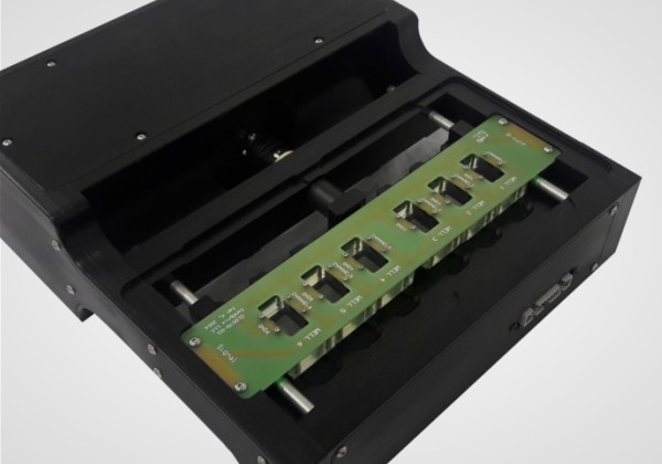
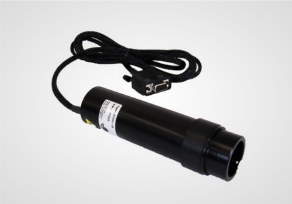
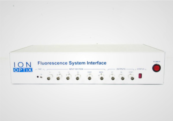
 under software control. PMT recording occurs at rates as high as 1000Hz through IonWizard and PMTACQ, our PMT module add-on. For stand-alone applications, a separate [...]

PMT Sub-System2019-11-11T10:37:43-05:00
Go to Top