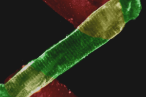
Muscle tissue provides many of the same important functional outputs as isolated myocytes while being easier to prep, more robust, and with added physiological relevance due to its multicellular context.
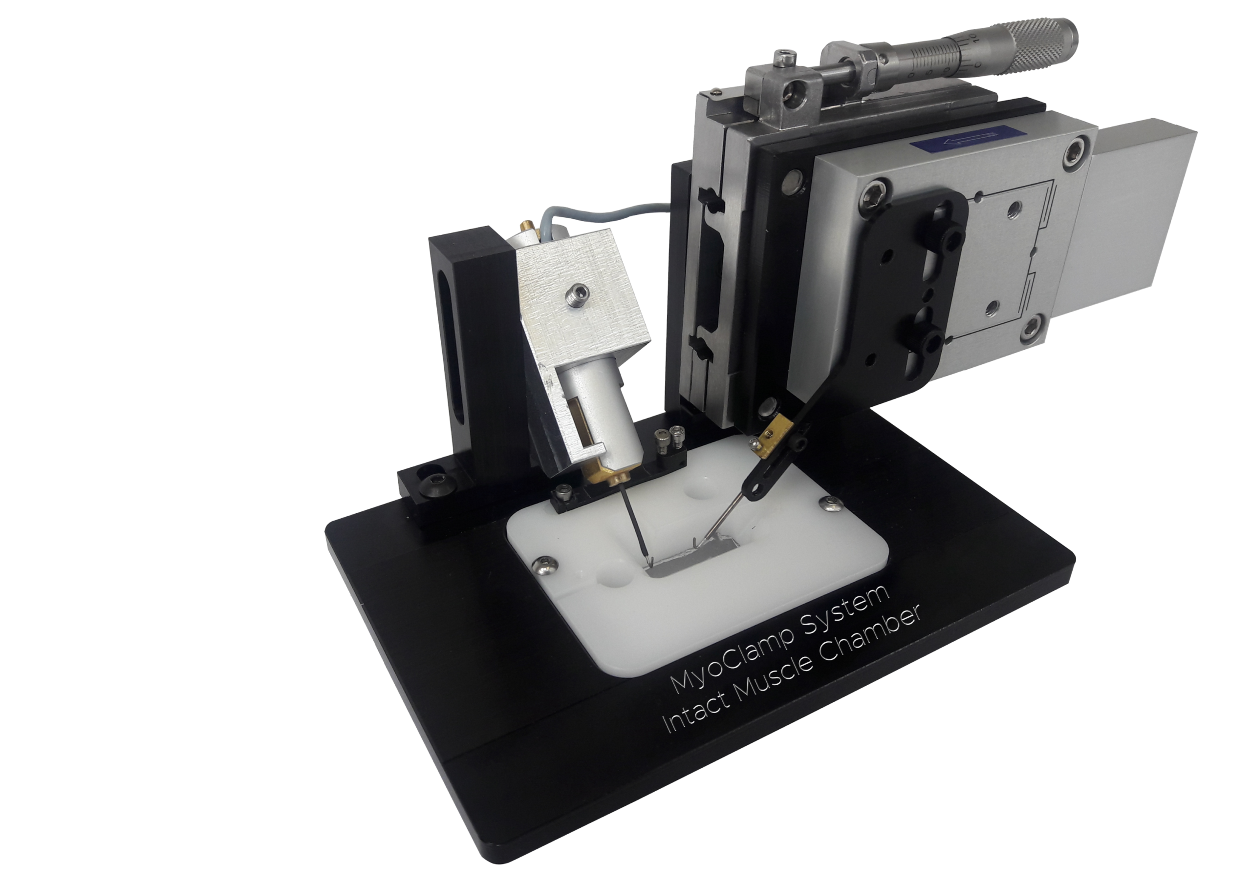
The IonOptix MyoClamp System provides high-content, functional data from multiple different tissue types including living myocardial slices, papillary muscle, and engineered heart tissue as well as skeletal preparations like soleus and EDL muscle. The system features interchangeable inserts to accommodate various tissue sizes, while also allowing tissue to be field stimulated via parallel platinum electrodes or stimulated via direct electrical excitation. Tissue can be easily affixed to sturdy, stable clips that mount between a robust force transducer and programmable length controller, or via simple platinum “Omega” clips tied to the muscle and slipped onto the platinum hooks of the force transducer and motor. The platinum used for electrical conductance are biologically inert and minimize electrolysis. The MyoClamp System’s Intact Muscle Chamber allows fluid flow for temperature control and continuous oxygenation of tissue, ensuring consistent and reliable data acquisition.
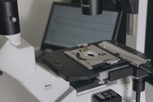
The platform can be used as a benchtop unit or, thanks to our chamber’s optically transparent bottom, mounted atop the stage of an inverted microscope for imaging. When combined with the IonOptix Calcium and Contractility System, fluorescence of quantitative, ratiometric dyes such as Fura-2 and/or CalRed can be detected using IonWizard, also permitting control of experiment acquisition parameters and comprehensive data analysis. Resting sarcomere length can also be detected in thin tissue preparations (a useful method to set cardiac tissue preload).
Our KoForce force transducer permits sensitive, high-speed, high-resolution acquisition of force development from as little as 15 nN to over 100 mN. Equipped with our robust moving coil long travel actuator with 20 nm resolution, the MyoClamp System enables force-feedback length control in cardiac tissue to emulate pressure-volume loops (requires minimum forces of ~1 mN). These force-length and stress-strain work loops can be used to derive important parameters using IonWizard’s Power Analysis to describe important functional characteristics including end-systolic and end-diastolic force-length/stress-strain relationships characterizing the mechanical stiffness of the tissue as well as contractile performance.
SPECIFICATIONS
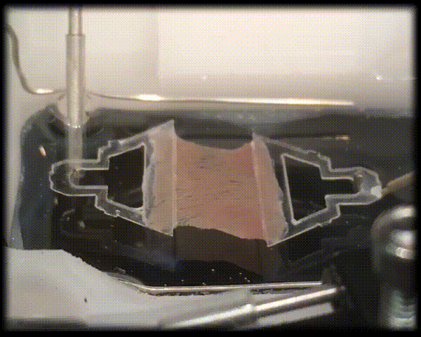
Cardiac tissue such as living myocardial slices, papillary muscle, and EHTs, unlike single isolated myocytes, can be used to study cardiac function within a multicellular context and an intact myofilament lattice. Cardiac tissue preparations have the added benefit of maintaining a syncytium of myocytes found in vivo thus maintaining in vivo architecture and intercellular signaling, suggesting that experimental results are more likely to have physiological relevance. And unlike whole heart studies, the contractile characteristics of these preparations can be evaluated independently of extrinsic factors such as vascular tone. These preparations also allow measurements too difficult or impossible to perform in whole hearts. And, unlike skinned preparations, intact muscle preparations allow for simultaneous detection of force production and intracellular calcium dynamics. Not limited to cardiac tissue, our MyoClamp System has been tested and used with a number of skeletal preparations including EDL, soleus, and engineered skeletal tissue.
The cardiac slice preparation technique has been recently championed by Professor Cesare Terracciano (Imperial College London) and his laboratory, including Dr. Fotios Pitoulis. We would like to acknowledge and thank them for their assistance with the tissue preparation.
We stand behind every product we sell, and we are committed to ensuring you get the most out of your IonOptix system. Our goal is to help you collect meaningful, reliable data as quickly as possible. Whether you need help with your system or have questions about the application, we’re here for you.


FEATURES
COMPONENTS
The IonOptix MyoClamp System can be combined with our typical Calcium and Contractility components to give a full suite of simultaneous fluorescence and contractility (i.e. force development) measurements. Depending on your specific application, systems can include a sensitive force transducer and/or a programmable length controller.
BROWSE THE TABS to learn more about the components that complete this system.

IonOptix-configured inverted microscopes provide an ideal platform for combined photometry and dimensioning measurements. These microscopes have been engineered to incorporate various unique IonOptix optical components and connectors in order to operate as the imaging component within our Calcium and Contractility Systems. Choose between the Motic AE31 or the Olympus IX73 depending on your application. LEARN MORE

The fluorescence excitation light source is chosen with your specific research needs in mind and can be configured for almost any dye or combination of dyes. Options include the ultra-fast HyperSwitch, monochromatic LEDs, or the more affordable MuStep. LEARN MORE

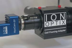
The sensors within the Intact Muscle Chamber system are responsible for translating cellular signal into data. Cell contractility is measured using the high-speed MyoCam-S3 digital camera, and whole-cell fluorescence photometry is measured with a photomultiplier tube (PMT). For cleaner fluorescence signals, the Cell Framing Adapter allows you to frame a cell of interest and reduce background fluorescence. LEARN MORE

The robust force transducer measures muscle force development with a typical sensitivity range of 0-100 mN. LEARN MORE

The field stimulator is an important component of excitation-contraction studies to provide acute stimulation of tissue. Use either the MyoPacer for simple stimulation protocols or the MyoPacer EP for advanced sequencing and arrhythmic stimulation. LEARN MORE

The specially-designed muscle chamber allows for simple mounting of muscle preparations, includes inlet and outlet for perfusion flow, and is compatible with almost any type of inverted microscope stage. LEARN MORE

The FSI ties the Calcium and Contractility system components together by communicating between hardware and the computer, allowing for synchronous control of contractility and/or calcium measurements as well as inputs and outputs for external devices. LEARN MORE

Several options are available to tailor programmable length control to your studies based on length range, resolution, and frequency response. LEARN MORE

The optional Campden Instruments 9000SMZ Vibrating Microtome delivers unmatched control, slice quality, and tissue viability — making it the ideal vibratome for living myocardial slice preparations. LEARN MORE
SOFTWARE
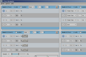
MyoClamp’s data acquisition is achieved through IonWizard, in which trace recordings display force data and length controller position among other potential data streams. Add software module PMTAcq for fluorescence recording, SarAcq for resting sarcomere length in thin striated tissue, Advanced Signal Generator for precise muscle length control with programmable protocols, and/or MultiClamp for force-length work loops.

RESOURCES
If you can’t find the information you are looking for, contact us!
